
This website uses cookies so that we can offer you the best possible user experience. The cookie information is stored in your browser and performs functions such as recognizing you when you return to our website or helping our team to understand which sections of the website you find most interesting and useful.
Neurocloud VOL

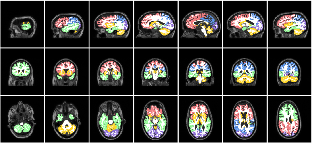
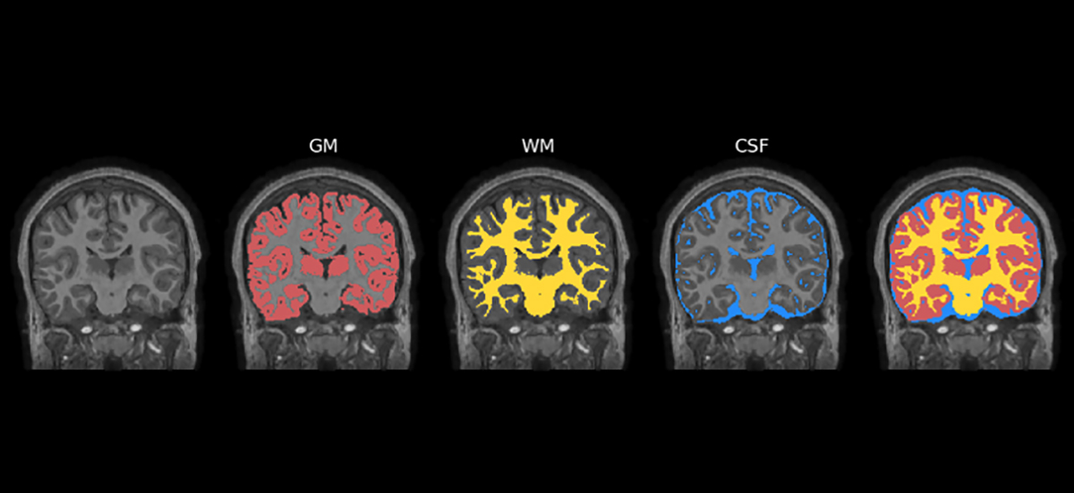
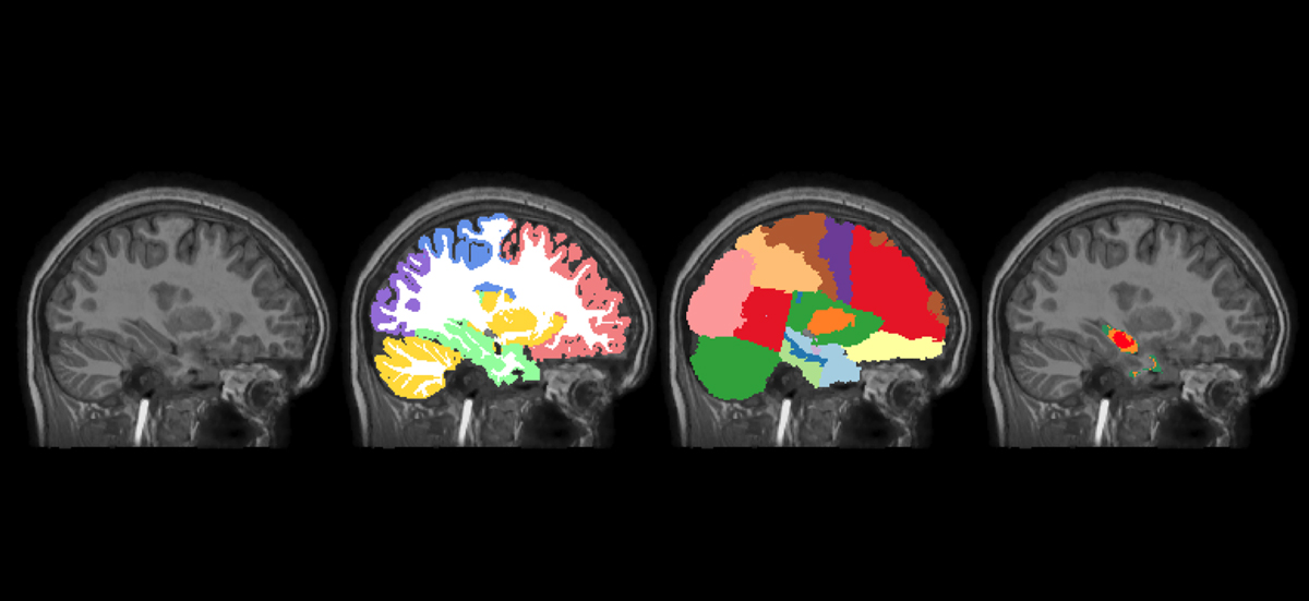
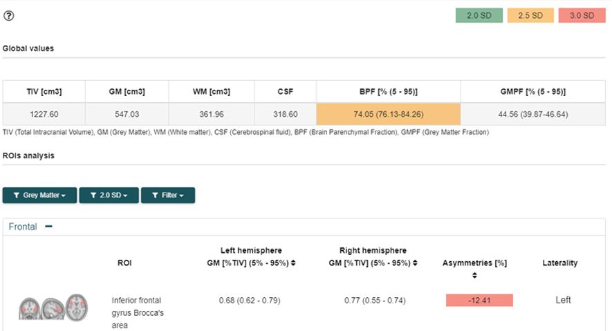
On the MRI T1 image, the software identifies atrophy in the patient’s brain. Cortical, subcortical, and other clinically relevant brain structures are segmented and volumetric data from the patient’s brain are compared with the normality distribution derived from a large age-stratified database of healthy controls. The results include data of interest such as BPF or GMPF, analysis by regions of interest, and voxel-to-voxel parametric analysis.
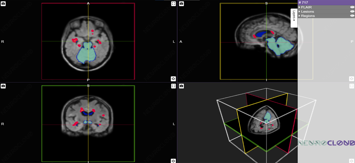
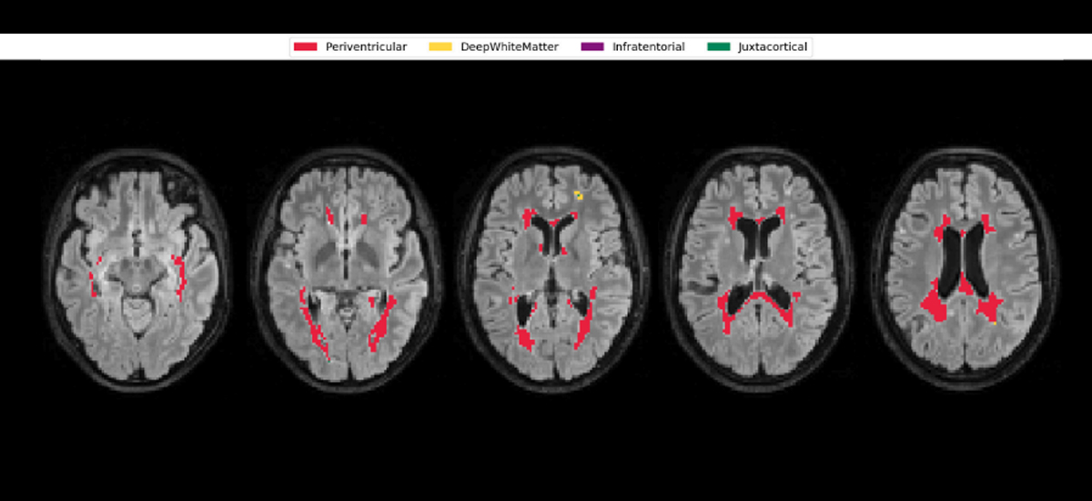
In the FLAIR image, the software segments and identifies inflammatory lesions that appear in the FLAIR image as hyperintense regions in white matter, and that are a pathological sign in the study of diseases such as sclerosis multiple. The results include the number and volume of segmented lesions, differentiating four classes of lesions: periventricular, subcortical, juxtacortical, and infratentorial.
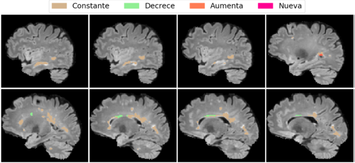
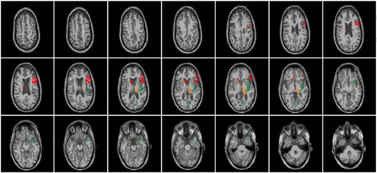
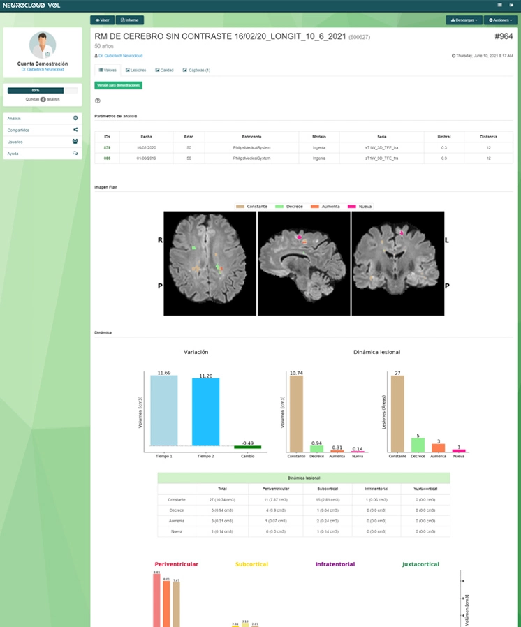
Neurocloud – VOL assists in the follow-up and evolution of the pathology, providing a longitudinal analysis of the imaging studies. The software presents in graphics and images the evolution of the atrophy and the growth / decrease of the lesions and the appearance of new lesions.

Help in radiology
Providing automatic segmentation of all brain areas and identification of atrophies and lesions. Help with visual analysis, confirm your findings, and improve your reports by adding quantitative information.
Help in neurology
Quantitative information allows you objective and personalized treatment and monitoring of the disease.
Help in diagnosis
Independent of the observer
Eliminates inter-observer differences between specialists. Enables you to move from a subjective working model to an objective model based on observer-independent data.
Automatic
Automatic processing and results in minutes. Connectivity with PACS systems for seamless integration with your workflow.
Sensitive
Identify atrophic VOI in the first stages of the disease thanks to one of the most extensive normal bases on the market, stratified by age.
All in one
It integrates all the necessary resources for the diagnosis:
results in images, data tables and interactive viewer.
Neurocloud VOL: Volumetric analysis for the quantification of atrophy and lesions in MR imaging
> Brain segmentation> Quantification of gray matter and white matter atrophies> Extraction of Z-scores and deviations from normality> Morphometric analysis based on voxel> Segmentation and quantification of lesions> Analysis of the evolution of lesions

100% Compatible with your equipment and PACS

Fast, intuitive and fully automated

Invisible integration in clinical practice

Continuous and free updates

Continuous and personalized assistance

Hiring adapted to your needs

Clinically validated, CE marked
